Parameter "weighting"
What is meant by a T1- or T2-weighted image?
|
|
Terms such as "T1-weighted" and "T2-weighted" are among the most overused and least understood concepts in MR imaging. In the broadest sense, these terms are used to communicate to other physicians the type of MR pulse sequence employed to generate a series of images. In a more narrow sense an implication exists that a single intrinsic tissue parameter (T1, T2, spin-density (ρ), diffusion, susceptibility, chemical shift, flow, perfusion, etc.) dominates the image contrast observed.
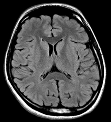 T2-FLAIR image
Medical students and non-radiologists are often taught to look at the the "color" of CSF or other fluids to determine the type of "weighting" — dark CSF means "T1-weighting" and bright CSF means "T2-weighting". Although this simple scheme may have worked 20 years ago, consider the brain image from the commonly used T2-FLAIR sequence pictured left. This sequence is known to have strong sensitivity to T2 changes, but the CSF signal has been suppressed by an inverting pulse and rendered black!
A fundamental misconception about "T1,T2, ρ, etc -weighting", is that contrast in the image is dominated by one specific tissue parameter to the exclusion of all others. To show the fallacy of this reasoning I developed an index system to quantify the relative contributions of these parameters on routine spin-echo brain imaging. The results were surprising, and showed among other things:
- Spin density (ρ) contrast is always present, contributing 30-50% of the contrast effect on both short TR/short TE (traditionally called "T1-weighted") and long TR/long TE ("T2-weighted) images.
- Short TR/short TE sequences often have as much "T2-weighting" as they do "T1-weighting".
- Long TR/long TE images do not have much "T1-weighting" but do have significant "ρ- weighting" in addition to "T2-weighting".
- The degree of "weighting" is not the same for all tissues in a given image.
|
Another common misconception is that T1-weighted or T2-weighted images are parameter "maps" whose pixel intensities are proportional to tissue T1 or T2 values. This is patently incorrect. Actual T1 and T2 maps are shown left so the reader may appreciate how different they appear compared to routine T1- or T2-weighted images.
|
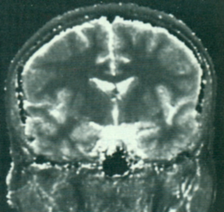
T1 map
|
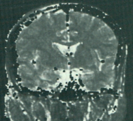
T2 map
|
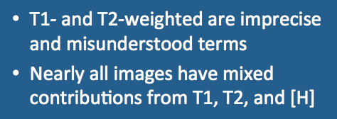
In summary, it is OK to continue to use terms like "T1-weighted" and "T2-weighted" as long as you realize they are imprecise, are not parameter "maps", and that nearly all images have mixed contributions from all the different tissue parameters (T1, T2, ρ, diffusion, susceptibility, chemical shift, flow, perfusion).
References
Elster AD. An index system for comparative parameter weighting in MR imaging. J Comput Assist Tomogr 1988; 12:130-4.
Ma YJ, Fan S, Shao H, Du J, Szeverenyi NM, Young IR, Bydder GM. Use of Multiplied, Added, Subtracted and/or FiTted Inversion Recovery (MASTIR) pulse sequences. Quant Imaging Med Surg 2020;10(6):1334-1369. [DOI LINK] (This paper applies the concept of Tissue Property Filters from the paper by Young et al below to understand more complex sequences such as the Double Inversion Recovery, MP2RAGE and various type of Subtracted Inversion Recovery methods -- a feat impossible to do in a useful way using the conventional qualitative concept of weighting.)
Ma YJ, Shao H, Fan S, Lu X, Du J, Young IR, Bydder GM. New options for increasing the sensitivity, specificity and scope of synergistic contrast magnetic resonance imaging (scMRI) using Multiplied, Added, Subtracted and/or FiTted (MASTIR) pulse sequences. Quant Imaging Med Surg 2020; 10:2030-2065. [DOI LINK] (This paper takes up the concept of Synergistic Contrast MRI (scMRI) where one tissue property is used twice or more in a single sequence to increase the contrast produced by it, or two or more different tissue properties are used in a single sequence for the same purpose.)
Yokoo T, Bae WC, Hamilton G, et al. A quantitative approach to sequence and image weighting. J Comput Assist Tomogr 2010;34:317-331.
Young IR, Szeverenyi NM, Du J, Bydder GM. Pulse sequences as tissue property filters (TP-filters): a way of understanding the signal, contrast and weighting of magnetic resonance images. Quant Imaging Med Surg 2020; 10:1080-1120. [DOI LINK] (An excellent review article, significantly expanding the work of Elster and Yokoo et al., formulating imaging contrast in terms of tissue-property filters and clarifying many paradoxes and misunderstandings concerning the concept of "weighting".)
Related Questions
I know long TR/TE gives T2-weighting and short TR/TE gives T1-weighting, but I don't understand why. Can you explain?
I know long TR/TE gives T2-weighting and short TR/TE gives T1-weighting, but I don't understand why. Can you explain?