MR Safety: Breast biopsyWhich breast biopsy devices and markers are MRI compatible?
|
|
Breast imaging specialists are often asked to place a wire, clip, or other marker to locate a site for future surgical biopsy or to document the site of a percutaneous biopsy. The vast majority of these localizations are done under mammography or ultrasound guidance, with MRI reserved only for lesions difficult to see on other modalities.
Most wires and markers in common use were developed before the widespread availability of breast MRI. As such, many have either not been formally tested by ASTM methods (though are likely of little risk to the patient because of their small size and deep locations). A devices few have been found to be "MR Unsafe", especially several types of biopsy guns and aspiration equipment. Fortunately, many new MR-compatible wire, marker and biopsy systems have been developed over the last two decades rendering most of these concerns moot.
Most wires and markers in common use were developed before the widespread availability of breast MRI. As such, many have either not been formally tested by ASTM methods (though are likely of little risk to the patient because of their small size and deep locations). A devices few have been found to be "MR Unsafe", especially several types of biopsy guns and aspiration equipment. Fortunately, many new MR-compatible wire, marker and biopsy systems have been developed over the last two decades rendering most of these concerns moot.
Wires for Breast Biopsy
Metallic wires, often ending with barbs or hooks, are commonly placed in suspicious lesions to guide the surgeon at open biopsy. These wires are typically placed on the morning of the planned surgery, and at the request of the surgeon, sometimes include the trocar or guiding needle in place taped to the skin. If not MRI compatible, these devices could conceivably cause heating or skin burns (like any juxtacutaneous metal object). But since patients nearly always go to surgery shortly after wire/hook placement, it is distinctly rare that they undergo MRI before the wire has been removed.
In the highly unusual case that one does encounter a patient with a protruding wire or needle, it is important to check the manufacturer's website to establish MR compatibility. Considerable care should be undertaken with exact reference to the manufacturer/wire name/diameter/length/SKU. For example, BD-Bard® currently sells 14 wires with exactly the same name (“Ghiatas™ Beaded Breast Biopsy Localization Wire”) but with different materials, lengths, diameters; only 4 of the 14 are MRI compatible!
In the highly unusual case that one does encounter a patient with a protruding wire or needle, it is important to check the manufacturer's website to establish MR compatibility. Considerable care should be undertaken with exact reference to the manufacturer/wire name/diameter/length/SKU. For example, BD-Bard® currently sells 14 wires with exactly the same name (“Ghiatas™ Beaded Breast Biopsy Localization Wire”) but with different materials, lengths, diameters; only 4 of the 14 are MRI compatible!
Biopsy Markers
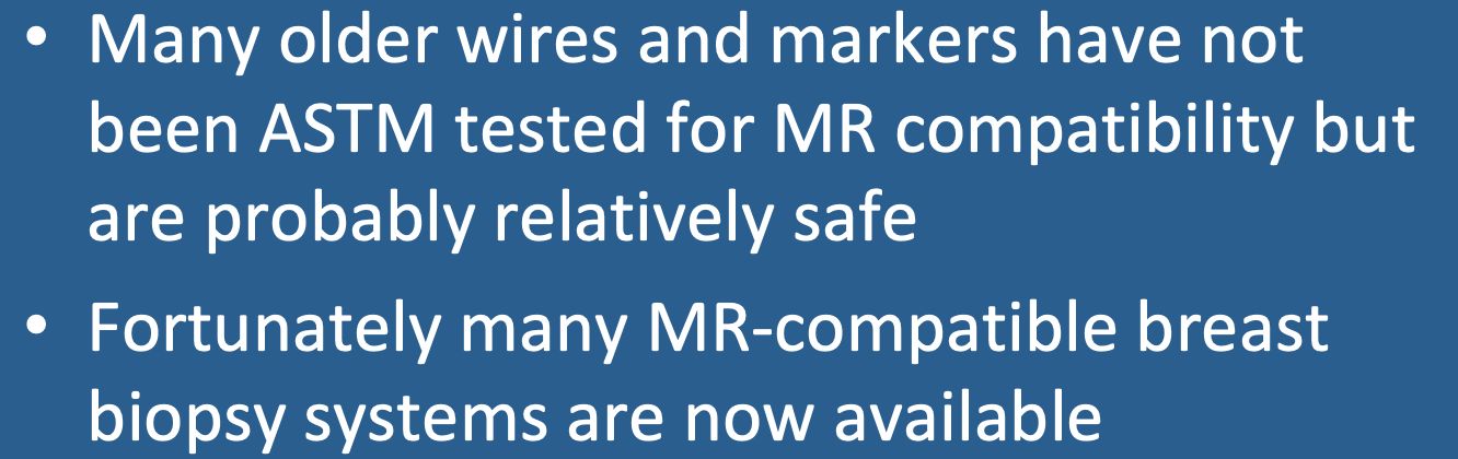
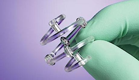
 Hologic® MR-compatible biopsy markers
A wide variety of MRI-compatible breast biopsy markers are now available. These are typically made of (nonferromagnetic) titanium, stainless steel, or other alloys. They are now commonly placed even in mammography or ultrasound so that future MRIs will not be impaired. If an unknown older marker composed of ferromagnetic steel is encountered, it would likely not be a safety issue, but could cause and appreciable susceptibility artifact on MRI.
|
In addition to the commonly used metallic markers, several new devices should be recognized that are unusual in nature and may be unfamiliar to most readers. Although placed under x-ray or ultrasound guidance (not MRI), patients with these markers may occasionally be referred for MRI. All are considered MR Conditional.
|
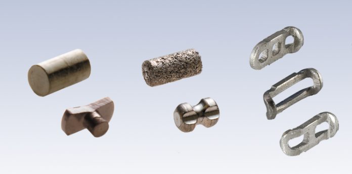
"MR Conditional" SAVI SCOUT® Radar Reflector Marker

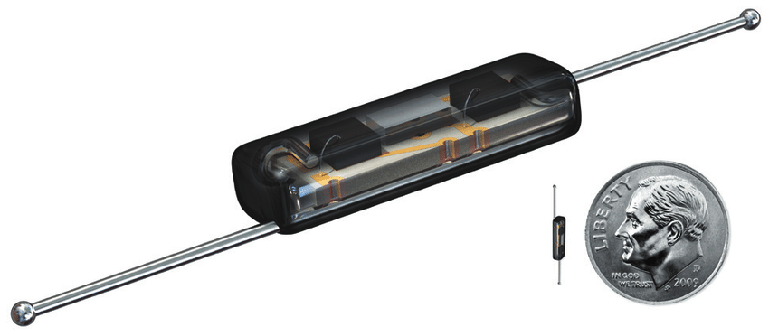
"MR Conditional" Magseed® ferromagnetic marker.
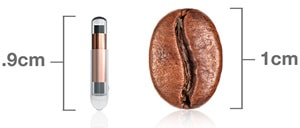
"MR conditional" LOCalizer™ RFID Tag
"MR Conditional" BioZorb® 3D marker.
|
References
Hayes MK. Update on preoperative breast localization. Radiol Clin N Am 2017; 55:591-603. [DOI LINK]
Kapoor MM, Patel MM, Scoggins ME. The wire and beyond: recent advances in breast imaging preoperative needle localization. RadioGraphics 2019; 39:1886-1906. [DOI LINK]
Portnow LH, Thornton CM, Milch HS, et al. Biopsy marker standardization: what’s in a name? AJR Am J Roentgenol 2019; 212:1400-1405. [DOI LINK]
Related Questions
What's the danger with breast tissue expanders in MRI?
What's the danger with breast tissue expanders in MRI?